Narendra Malhotra (Doctor)*
Jaideep Malhotra (Doctor)*
Pranay Shah
Vanaj Mathur
Nidhi Gupta
Amita Samiksha
Malhotra Nursing and Maternity Home, Agra, India
TThe patient, a 28-year-old woman, presented with secondary infertility of 5 years duration. Her first child (a daughter now aged 5 years) was delivered normally. At presentation, she complained only of dysmenorrhoea, lower abdominal pain, and backache.
Various diagnostic tests were performed. Screening colour doppler sonosalpingography revealed patent fallopian tubes, a normal uterus, and a normal endometrial canal. However, both ovaries were enlarged and each contained a small hyperechoric mass: 1.6 ¥ 1.6 cm in the right ovary, and 2.5 ¥ 2.5 cm in the left ovary (figures 1, 2).
X-ray of the lumbar spine was inconclusive, but magnetic resonance imaging of the pelvis (see figure 3) revealed the following:
1. Lobulated space-occupying lesion (SOL) in the left ovary suggestive of lipomatous tissue. 2. Focal lesion in the right ovary of hypo- to hyper-intensity on T1- and T2-weighted sequences.
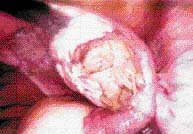
Diagnostic laparoscopy (figures 4, 5) confirmed the enlarged ovaries. The SOL was removed (cystectomy). Tubal patency was established.
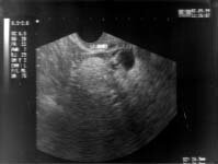 |
| figure 1. |
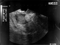 |
| figure 2. |
|
|
- What is the most likely diagnosis?
- What are the ultrasound characteristics of this condition?
- How does it cause infertility?
- What is the ideal management approach (and what precautions should be taken)?
- What are the prospects for restoring fertility?

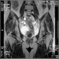 |
| figure 3. |
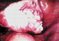 |
| figure 4. |
 |
| figure 5. |
|