|
|
||||||||||||||||||||||||
|
||||||||||||||||||||||||
| Table 1. Age distribution of patients with carcinoma of the cervix. |
 |
| Table 2. Presenting complaints of patients with carcinoma of the cervix. |
 |
| Table 3. Duration of disease. |
 |
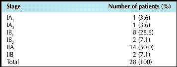
Of the 28 patients, 26 had squamous cell carcinoma (92.9%) and 2 had adenocarcinoma. According to the 1994 International Federation of Gynecology and Obstetrics (FIGO) clinicopathological staging system, the largest group of patients was those with stage IIA disease, accounting for 50% of all patients (Table 4). Seven of 28 patients had lymphatic spread (25%). The number of dissected nodes ranged from 8.0 to 44.0 (mean, 21.6). The resected vaginal margins were divided into 3 categories according to the postoperative histopathological evaluation of the specimen (Table 5).
| Table 4. Clinicopathological staging of disease. |
 |
| Table 5. Status of resected margins of the vagina. | ||||||
 |
||||||
|
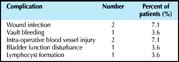
Postoperative drainage was 100 ml to 500 ml (average, 350 ml) and the duration of drainage was 48 to 72 hours. Five patients developed a total of 7 intra-operative or postoperative complications, accounting for 17.9% of the 28 patients (Table 6). There was no postoperative mortality. Eight patients received postoperative radiotherapy. Five of these patients had lymph node metastases, 2 had parametrial infiltration, and 1 had a positive margin at the vaginal vault.
| Table 6. Intra-operative or postoperative complications. |
 |
Discussion

Worldwide, the incidence of carcinoma of the cervix is decreasing. However, carcinoma of the cervix is still a major malignancy endangering female health in relatively underdeveloped countries. According to the statistics of 45 countries conducted in 2000, the incidence of cervical carcinoma is high in Mexico 17.1/100,000, Venezuela 15.2/100,000, and Tobago 15.0/100,000.3 Even though Nepal does not have its own data on the incidence or prevalence of cervical carcinoma; early marriage, early childbearing, and multiparity predispose women to the disease in this country.
In a study of histopathological specimens at the BP Koirala Memorial Cancer Hospital from July 1999 to January 2001, 272 cases of cervical carcinoma diagnosed accounted for 84.73% of 321 gynaecological malignancies and 31.16% of all 873 cancers.4 The highest incidence was among the 31 to 50 years age group. This is in concordance with our observation that the median age was 44 years.
Surgical therapy and radiotherapy are the 2 main treatment modalities for carcinoma of the cervix. For stage 0 and IA disease, conservative surgery is sufficient. For patients with stage IIB to IV disease, radiotherapy and/or chemotherapy is the standard choice of treatment. For stage IB and IIA disease, radical surgery or radiotherapy and/or chemotherapy are equally effective with overall 5-year survival rates of 80% to 90%. Landoni et al performed the only prospective randomised trial for patients with stage IB or IIA disease to compare radical surgery to radiotherapy.5 The 5-year actuarial disease-free survival rates for patients with tumours 4 cm or smaller (stage 1B![]() ) treated with surgery or radiotherapy were 80% and 82%, respectively. Disease-free survival was 63% and 57%, respectively, for patients with larger tumours. Radiotherapy with concurrent chemotherapy (cisplatinum 40 mg/m2 and 5-flurouracil 1000 mg/m2) is the standard of care for locally advanced cancers of cervix.6-8
) treated with surgery or radiotherapy were 80% and 82%, respectively. Disease-free survival was 63% and 57%, respectively, for patients with larger tumours. Radiotherapy with concurrent chemotherapy (cisplatinum 40 mg/m2 and 5-flurouracil 1000 mg/m2) is the standard of care for locally advanced cancers of cervix.6-8
The radiation field for carcinoma of the cervix consists of pelvic lymph drainage areas and para-aortic drainage areas. Some authors advocate irradiation to both areas as a prophylactic measure. Rotman et al divided 337 patients with stage IB and IIA disease into 2 groups to receive standard radiotherapy or standard radiotherapy plus para-aortic lymph node irradiation The 5-year survival rate was 55% and 67%, respectively, indicating a survival benefit for the latter group of patients.9 However, irradiation of the large fields will inevitably bring more morbidity. For stage IB and IIA disease, the rate of pelvic lymph node metastasis is low (4.3% to 34.0%) in this series it was 25%. If only patients with stage IB, IIA, and IIB disease were considered, the rate was 26.9%. Therefore, information about postoperative lymphatic metastases will provide the reference for delivery of radiotherapy and prevent unnecessary morbidity.
To a great extent, the choice of treatment is primarily based on a patient's preference, availability of treatment facility, anaesthetic and surgical risks, and physician's preference. In the context of Nepalese patients, it is considered that patients with stage IB and IIA disease will derive most benefit from surgical therapy, for the following reasons: surgical expertise is readily available; radiotherapy machines are not readily available; for surgical therapy, the treatment period is short and cheap, which is readily accepted by patients of a poorer socioeconomic background; and the prevalent age of patients with cervical carcinoma is approximately 40 years. Surgical therapy will preserve functional ovaries and prevent stenosis of the vagina, thereby maintaining quality of life. Moreover excision shows the exact lymph node status, decreasing the size of the radiation field and consequent morbidity.
Effective pelvic drainage prevents pelvic infections, vaginal stump fistulas, and lymphocele. Rational use of pelvic drainage can decrease the complication rate. In this group of patients, the vaginal glove drain gave satisfactory results, with average drainage of 350 ml, and an average drainage time of 48 to 72 hours. No pelvic infection or vaginal fistulas were noted and only 1 patient (3.6%) had lymphocele, similar to the incidence of <5% reported in the literature for retroperitoneal suction drainage.8 Furthermore, compared with the standard drain tube, this method is easy to place, drains freely, and does not require scalpel wounds in the lower abdomen, resulting in reduced ambulation.
Conclusion

Surgical excision is a primary therapy for early stage carcinoma of the cervix. Although radiotherapy and/or concurrent chemotherapy results in equivalent survival, surgery is preferred for this subset of patients. The vaginal glove is a safe, effective, and simple method of pelvic drainage.
References

-
Parkin DM, Pisani P, Ferlay J. Estimates of the worldwide incidence of 25 major cancer in 1990. Int J Cancer 1999;80:827-841.
-
Eifel PI, Berek JS, Thigpen JT. Cancer of the cervix, vagina, and vulva. In: DeVita VT, Hellman S, Rosenberg SA, editors. Cancer: principles and practice of oncology. 6th ed. New York: Lippincott Williams and Wilkins; 2001:1526-1573.
-
Jemal A, Thomas A, Murray T, Thun M. Cancer statistics, 2002. CA Cancer J Clin 2002;52:23-47.
-
Pradhan M, Dhakal HP, Pun CB, et al. Gynecological malignancy in BPKMCH, Bharatpur: a retrospective analysis of 321 cases. J Nepal Med Assoc 2001;40:108-111.
-
Landoni F, Maneo A, Colombo A, et al. Randomised study of radical surgery versus radiotherapy for stage Ib-IIa cervical cancer. Lancet 1997;350:535-540.
-
Rose PG, Bundy BN, Watkins EB, et al. Concurrent cisplatin based radiotherapy and chemotherapy for locally advanced cervical cancer. N Engl J Med 1999:340;1144-1153.
-
Morris M, Eifel PJ, Lu JD, et al. Pelvic radiation with concurrent chemotherapy compared with pelvic and para-aortic radiation for high risk cervical cancers. N Engl J Med 1999;340:1137-1143.
-
Rotman M, Pajak M, Choi K, et al. Prophylactic extended field irradiation of para-aortic lymph nodes in stages IIB and bulk IB and IIA cervical carcinomas: ten-year treatment results of RTOG 79-20. JAMA 1995;274:387-393.
- Shinglelon HM, Thompson JD. Cancer of the cervix. In: Rock JA, Thompson JD, editors. Operative gynecology. 8th ed. New York: Lippincott-Raven; 1997:1413-1499.
|
Addendum
|
|
Intracavitary radiotherapy became possible at |
|
Address for Correspondence
|
|
Dr Nirmal Lamichhane |
