
Cavitating Lung Lesion in a Patient with Neutropenic FeverA 43-year-old woman presented with fever, bleeding tendency, and a high white cell count of 163 x 109/L (normal range, 4.5 to 11.0 x 10⁹/L). Subsequent investigations confirmed a diagnosis of Philadelphia chromosome-positive acute lymphoblastic leukaemia. She received 2 courses of combination chemotherapy and achieved partial response. Thereafter she received 2 further courses of salvage combination chemotherapy; however, the response was poor with residual leukaemia.
|
|||||
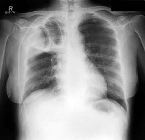
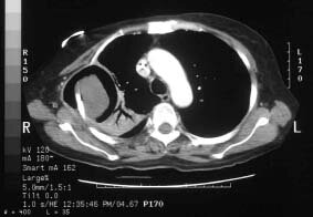
Figure 2. |
Computed tomography of the thorax showing a 6 x 6 x 6 cm non-calcified lesion involving the lateral aspect of the right upper ribs with extra-thoracic extension. The lesion was cavitatory with a relatively smooth border and intraluminal roundish lesions that gave rise to the crescent sign in keeping with aspergilloma |
The lesion was cavitatory with a relatively smooth border and intraluminal roundish lesions that gave rise to the crescent sign, which is in keeping with pulmonary aspergilloma. Associated peri-lesional consolidation was noted (Figure 2). In view of the persistent myelosuppression, further invasive investigation was withheld. Although the fungal culture for sputum was negative, the appearances of her chest radiograph and CT of the thorax, together with the fact that her fever responded to amphotericin, were consistent with the diagnosis of fungal chest infection.
Wing M Ho
Medical Officer
Department of Clinical Oncology
Prince of Wales Hospital
Shatin, NT
Hong Kong
China
![]()
Home | Current Issue | Back Issues | Congress Calendar
Free Subscription | Editorial Board |