Original Article

Click for Home

Atlas of Oculoplastic Surgery

J Junceda
Department of Ophthalmology, Hospital Valle del
Nalon, Asturias, Spain


Correction of Palpebral Ptosis via a Conjunctival Approach


Figure 1. Palpebral conjunctiva and tarsus transected. The orbicular muscle is grasped by Berke forceps.

Figure 2. The orbicular is freed from the forceps.

Figure 3. The anterior surface of the elevator fascia is dissected.

Figure 4. The captured tissues are assessed and the amount of fascia to be resected is measured.

Figure 5. The selected tissue is transected.

Figure 6. Samples for pathology examination.

Figure 7. Mild congenital palpebral ptosis seen preoperatively in the right eye.

Figure 8. The same patient after surgery.

Figure 9. Moderate to severe congenital palpebral ptosis seen preoperatively.

Figure 10. The same patient 1 month after surgery.

Figure 11. The same patient 6 months after surgery.

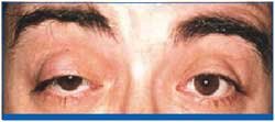
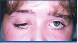
This Pictorial Oncology was submitted by Dr Juan Junceda and is based on a CD-ROM of 15 years experience of oculoplastic surgery. The CD-ROM contains more than 2 and a half hours of in vivo surgery and 32 films distributed in 5 sections of anatomy, palpebral surgery, conjunctival surgery, lacrimal pathway surgery, and orbital surgery.

The CD-ROM is currently distributed only in Spanish-speaking countries, although it will soon be edited by an International Publisher, which will make it more readily available.


Back to Top


Home - Table of Contents - Current Issue - Back Issues -
Congress Calendar
- Editorial Board
- - Free Subscription