|
Pearls in Glaucoma Filtration Surgery
A Euswas
Department of Ophthalmology, Ramathibodi Hospital, Faculty of Medicine, Mahidol University, Bangkok, Thailand
|
Trabeculectomy is a filtering procedure designed to direct the aqueous humor from the anterior chamber through a fistula to a subconjunctival reservoir, the filtering bleb. The goal of glaucoma filtration surgery (GFS) is to lower the intraocular pressure (IOP) below the threshold that causes optic disc damage in patients with uncontrolled glaucoma despite maximum tolerable medical therapy and who have a reasonably good prognosis for surgical success. Recently, some studies have supported performing trabeculectomy earlier in the course of glaucoma, even as primary therapy, but this concept is still under study and not generally acceptable.
This article describes the authors' techniques of standard GFS, step-by-step, emphasising the important surgical pearls for ultimate success.
Bridle Suture
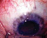
First, to stabilise the globe and to expose the surgical area, a bridle suture is placed approximately 9 to 10 mm posterior to the limbus. If topical or subconjunctival anaesthesia is used, a corneal traction suture will be placed into the clear cornea just anterior to the conjunctival insertion instead of a bridle suture (figure 1).
Figure 1. Corneal traction suture.

Conjunctival Incision
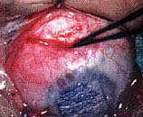
For a limbus-based trabeculectomy, the initial incision through the conjunctiva is performed approximately 9 to 10 mm posterior to the limbus (figure 2). I prefer the supero-nasal quadrant just nasal to the vertical meridian and leave the superotemporal quadrant available for possible subsequent filtering or cataract surgery.
Figure 2. Conjunctival incision.

If the patient will be undergoing posterior segment intervention in the near future, the surgical site should be at the 12 o'clock position to avoid sclerostomy ports and buckling procedure.
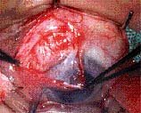
The initial incision through the conjunctiva is important. The dissection of the conjunctiva should be made 9 to 10 mm posterior and parallel to the limbus. The conjunctiva is dissected from the underlying Tenon's capsule towards the limbus. A small incision is made through the Tenon's capsule to the level of the episclera, 4 to 5 mm posterior to the limbus. Both blades of the scissors may be inserted into this small incision to the sub-Tenon's capsule space. The scissors blades are then rotated to a plane perpendicular to the globe and slightly separated to lift the conjunctiva/ Tenon's capsule flap away from the underlying episclera in both directions until the entire conjunctival insertion is reached (figure 3).
Figure 3. Conjunctiva/Tenon's capsule flap

The Tenon's capsule insertion will be encountered just at the posterior portion of the surgical limbus. The conjunctiva needs to be reflected anteriorly at least to this insertion. The light adhesions in this area can be lysed with a blunt-tipped small blade held perpendicular to the surface of the globe parallel to the limbus, advancing the blade sideways towards the limbus.
Half-thickness Scleral Flap
A small amount of cautery should then be applied to the limbal area to achieve haemostasis and to prepare for the subsequent preparation of the half-thickness flap. Various shaped flaps can be employed, including a trapezoid, triangular, square, rounded, or arch shape, each of half scleral thickness. I prefer the triangular flap and outline an area of the flap by using gentle bipolar or unipolar cautery.

 
|
